Ovarian Reserve Testing
Fertility Estimation (ovarian reserve testing)
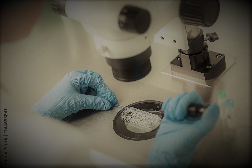
Compared to a healthy man whose sex cells (sperm) are created all of their lives, healthy women are already born with all the egg cells they will ever have. After birth, a woman’s egg number can only be reduced in puberty; it drops down from 6 to 7 million (number of egg cells in a fetus) to around 300.000. Thus, clinically significant fertility decline starts already in the early thirties. Each of approximately 400 menstrual cycles in a woman’s lifetime uses around 1.000 eggs to choose a single egg that will ovulate in that cycle. This happens due to a complex mechanism of choosing that one egg. Apart from this, other factors can influence the number and quality of a woman’s eggs. They are usually found in genetics, lifestyle, environment, or medical conditions such as endometriosis, ovarian surgery, chemotherapy, and radiation. All of this eventually leads to complete non-responsiveness of the ovaries and menopause. Ovarian reserve denotes the ability of ovaries to create eggs that, when fertilized, resulting in a healthy and successful pregnancy. Nowadays, we have tests telling us of the ovarian reserve. These tests use the information on the number and quality of the remaining eggs. Different tests can be done separately but only combined; they provide complete information on a woman’s reproductive potential. An ovarian reserve test in Iram clinic includes the following:
Biochemical tests:
Follicle-stimulating hormone (FSH)
Anti-Mullerian hormone (AMH)
Ultrasound:
antral follicle count (AFC)Follicle-stimulating hormone (FSH)
FSH is a hormone synthesized and secreted by the anterior pituitary gland situated in the brain. It stimulates the growth and maturing of ovarian follicles resulting in a mature egg ready to ovulate and be fertilized. FSH is secreted throughout a woman’s life, but in non-responsive ovaries, it can no longer stimulate follicular growth, and as a result, FSH levels increase. During menstruation, FSH stimulates the ovary to produce follicles. If the ovary no longer has follicles capable of growing, the FSH value will increase, making the ovary react by overstimulating it. For this reason, the FSH value is at its peak during the menstrual period, which is the optimal time for testing (day 2-5 of the period). Increased basal FSH value (higher than 10-20IU) indicates reduced ovarian reserve, resulting in an inadequate response to possible ovulation stimulation.
Anti-Mullerian hormone (AMH)
AMH is a glycoprotein, which in women is a product of granulose cells of primary, preantral, and antral follicles in the ovary. Therefore, AMH values directly depend on the number of remaining follicles in the ovary. Due to small follicles that secrete AMH, its concentration is stable and can be measured throughout the entire cycle. As AFC (the number of follicles) decreases with age, it causes a lowering of AMH levels, which is one of the indicators of the remaining ovarian reserve. In IVF procedures, AMH is linked with the number of retrieved eggs. High AMH suggests women will overreact to stimulation and have ovarian hyperstimulation syndrome (OHSS), and low AMH suggests a woman will have a poor response to ovulation stimulation. AMH levels are a good indicator of the quantity and number of eggs, but not of their quality. Therefore, a young woman with a low AMH level may have a reduced egg number but good egg quality with relatively good chances of getting pregnant. AMH values as fertility indicators in women under the age of 38 are approximate: AMH (pmol/L) 0.0-2.2 Very low concentration 2.2-15.7 Reduced fertility 15.7-28.6 Satisfactory fertility 28.6-48.5 Optimal fertility >48.5 Increased concentration Many labs will not use the table above but will read AMH levels according to age-connected reference values, creating false security in women. For example, in older women, reference values are narrowed so that an AMH result of 20 pmol/l in a 40-year-old woman, whose reference values are 1-25 pmol/l), represents a far better result than in a young woman, whose reference values range from 1 to 75 pmol/l. However, chances for pregnancy are better in younger women. Laboratories use different units to express AMH levels. If your result is expressed in μg/L, you can use the following formula to get a pmol/l result: x (level on the test result) x 7,14 = level in pmol/L Lately, it has been found that the AMH levels in a population of women are similar in women of the same age, i.e., an equal number of women with low AMH can be found in women of the same age in both fertile infertile populations of women.
Antral follicle count – AFC
Transvaginal ultrasound is used to count antral follicles 2-10 mm in diameter on both ovaries. The number of antral follicles directly indicates ovarian reserve. An infertility specialist can predict ovarian response to ovulation stimulation and the probability of the number of eggs retrieved in an IVF procedure based on AFC. Total number of antral follicles (expected ovulation stimulation medication response and success probability) < 4 (Extremely low count, poor response to stimulation. Possible canceling the procedure) 4 – 7 (Low count, probably a poor response to stimulation. High doses of gonadotropins needed) 8 – 10 (Moderately low count, slightly reduced chances of pregnancy) 11 – 14 (Normal count, good response to stimulation, good chances of pregnancy) 15 – 26 (Excellent response to mild stimulation, best number of expected pregnancies) > 26 (High count, very high risk of OHSS, and high probability of pregnancy) Biochemical tests and ultrasound can be used to estimate ovarian reserve. Since no ovarian reserve estimation has 100% sensitivity or specificity, tests are combined in order to get more precise results. AMH and AFC are the best indicators. Other tests such as estradiol and inhibin B levels, clomiphene citrate challenge test or ultrasound measurement of ovarian volume do not exert enough sensitivity and specificity and are no longer recommended for ovarian reserve estimation. Ovarian reserve estimation (testing) is recommended: – to women planning to postpone pregnancy beyond the age of 30 or 35, – to all women in ART procedures, – for genetic reasons (e.g., 45X mosaicism), – to women with a family history of premature ovarian insufficiency, – to women who may have ovarian damage (endometriosis, pelvic inflammatory disease, cyst, ovary removal, results of gonadotoxic medication treatment and/or pelvic carcinoma irradiation), – to women smokers. The primary goal of these tests is to identify women with a low ovarian reserve and then adjust treatment accordingly to all women who need treatment. We would like to stress that ovarian reserve testing only indirectly indicates fertility reserve in women. Young women with a low ovarian reserve can be entirely fertile, while women in their forties with a high ovarian reserve may be less fertile.


.jpg)


.jpg)

